|
НОС [ nose ] Нос - это орган головы, состоящий из наружного носа и, находящейся внутри него, полости носа.
НОС: ОГЛАВЛЕНИЕ
- НОС – nose
- ПОЛОСТИ НОСА – nasal cavities
- НАРУЖНЫЙ НОС – external nose
- СКЕЛЕТ НОСА – skeleton of the nose
- МЯГКИЕ ТКАНИ НОСА – soft tissue of the nose
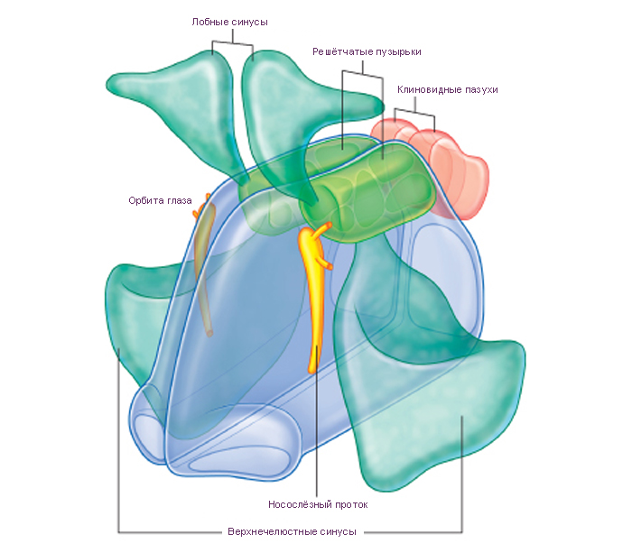
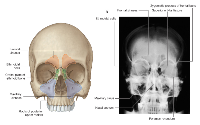
- ОКОЛОНОСОВЫЕ ПАЗУХИ – paranasal sinuses
- СТЕНКИ НОСОВЫХ ПОЛОСТЕЙ – walls of nasal cavities
- КАНАЛЫ НОСОВЫХ ПОЛОСТЕЙ – gateways to the nasal cavities
- КРОВОСНАБЖЕНИЕ НОСА – vascular supply of nasal cavities
- ИННЕРВАЦИЯ НОСОВЫХ ПОЛОСТЕЙ – innervation of nasal cavities
- ЛИМФАТИЧЕСКИЕ СТРУКТУРЫ НОСА – lymphatics of nasal cavities
 См. текст и иллюстрации ниже на странице. См. текст и иллюстрации ниже на странице.
- ФУНКЦИИ НОСА – functions of the nose
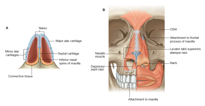
Наружный нос разделяют на следующие взаимосвязанные части: корень носа, спинка носа, верхушка носа и крылья носа.
Корень носа расположен в верхней части лица и отделен от лба выемкой, называемой переносьем. Боковые стороны наружного носа соединяются по срединной линии и образуют спинку носа. Нижние части боковых сторон наружного носа называют крыльями носа. Дистально спинка наружного носа переходит в верхушку носа. Крылья носа своими нижними краями ограничивают отверстия - ноздри носа. Они служат для прохождения воздуха в полость носа и из полости носа. Ноздри отделяются друг от друга подвижной (перепончатой) частью перегородки носа.
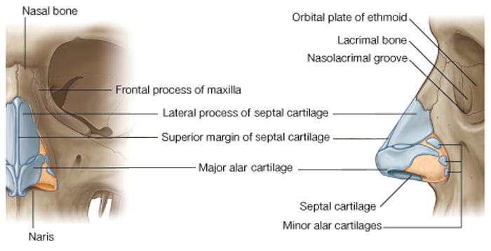
Наружный нос имеет костный и хрящевой скелет. Костный скелет образован носовыми костями и лобными отростками верхней челюсти. Хрящевой скелет образован несколькими гиалиновыми хрящами (остатки хрящевой носовой капсулы зародыша). Корень носа, верхняя часть спинки носа и боковых сторон наружного носа имеют костный скелет. Средняя и нижняя части спинки носа и боковых сторон носа имеют хрящевой скелет.
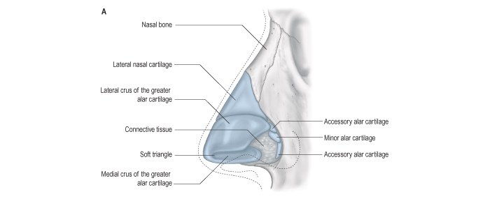
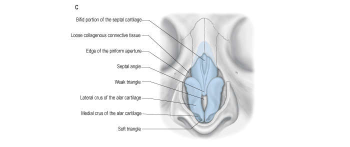
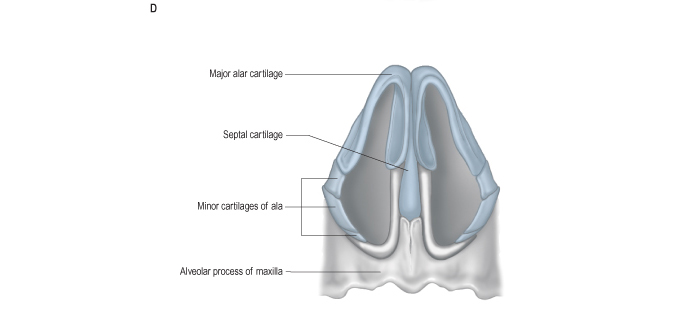
Латеральный хрящ носа - это парная треугольная структура, расположенная непосредственно ниже носовых костей. Латеральный хрящ носа принимает участие в образовании боковой стенки наружного носа. Передние края правого и левого боковых хрящей, соединяются между собой по срединной линии. Нередко срастаясь, они образуют единую структуру - спинку носа. Дистально (кпереди) латеральный хрящ с каждой стороны соединяется с большим хрящом крыла носа. Проксимально (сзади) латеральный хрящ прикреплен к нижнему краю носовой кости и к лобному отростку верхней челюсти.
Большой хрящ крыла носа - это парная структура, расположенная ниже соответствующего латерального хряща носа. Большой хрящ крыла носа ограничивает спереди и сбоку вход в полость носа и участвует в образовании ноздрей.
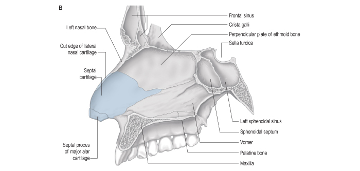
Малые хрящи крыла носа, два - три с каждой стороны, расположены позади большого хряща крыла носа, между ним и краем грушевидного отверстия. Иногда существуют несколько добавочных носовых хрящей. Эти различные по величине структуры расположены между латеральным хрящом носа и большим хрящом крыла носа. Изнутри, со стороны полости носа, к внутренней поверхности спинки носа примыкает край хряща перегородки носа. Хрящ перегородки носa, непарная структура, имеет четырехугольную форму и образует большую переднюю часть перегородки носа. Сзади и сверху хрящ перегородки носа соединяется с перпендикулярной пластинкой решетчатой кости. Сзади и снизу хрящ перегородки носа соединяется с сошником и с передней носовой остью. Между нижним краем хряща перегородки носа и передним краем сошника на каждой стороне располагается узкая полоска сошниково-носового хряща. Хрящи носа соединяются между собой и с прилежащими костями соединительной тканью.
Полости носа - это внутреннее пространство, образованное структурами наружного носа. Полость носа разделена перегородкой носа на две приблизительно симметричные ёмкости, которые открываются спереди на лице ноздрями. Сзади эти ёмкости через хоаны сообщаются с носовой частью глотки.
Перегородка носа - это спереди перепончатая и хрящевая структура, а сзади - костная структура. Перепончатая и хрящевая структуры вместе образуют подвижную часть перегородки носа.
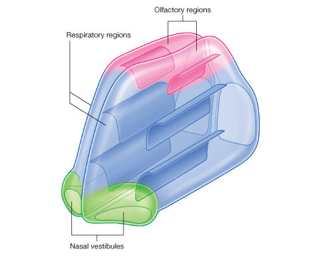
В каждой половине полости носа выделяют преддверие носа. Каждое преддверие носа сверху ограничено небольшим возвышением - порогом полости носа. Порог полости носа образован верхним краем большого хряща крыла носа. Преддверия носа покрыты изнутри продолжающейся сюда через ноздри кожей наружного носа. Кожа преддверий содержит сальные,потовые железы и жёсткие волосы - вибрисы.
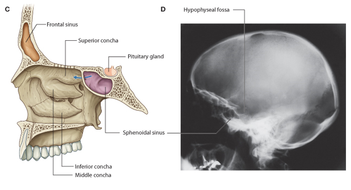
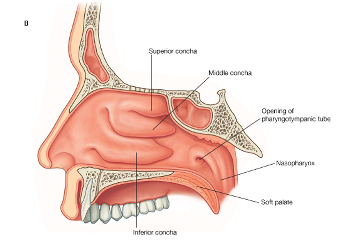
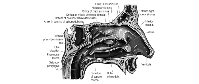
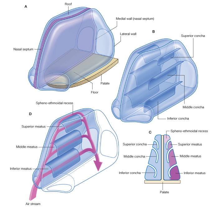
Большая часть полости носа представлена носовыми ходами. С носовыми ходами сообщаются околоносовые пазухи. Различают верхний носовой ход, средний носовой ход и нижний носовой ход. Каждый из них располагается под одноименной (верхний, средней и нижней) носовой раковиной. Позади и сверху от верхней носовой раковины находится клиновидно-решетчатое углубление. Между перегородкой носа и медиальными поверхностями носовых раковин расположен общий носовой ход, имеющий вид узкой вертикальной щели. Отверстие клиновидной пазухи находится в области клиновидно-решетчатого углубления. В верхний носовой ход открываются одним или несколькими отверстиями задние ячейки решетчатой кости. Боковая стенка среднего носового хода образует закругленное выпячивание в сторону носовой раковины - большой решётчатый пузырёк (выступающие средние ячейки решетчатой кости). Спереди и снизу большого решетчатого пузырька имеется глубокая полулунная расщелина. В её передней области находится нижний конец решетчатой воронки, через которую лобная пазуха сообщается со средним носовым ходом. Средние и передние ячейки (пазухи) решетчатой кости, лобная пазуха, верхнечелюстная пазуха открываются в средний носовой ход. В нижний носовой ход ведет нижнее отверстие носослезного протока.
Слизистая оболочка носа продолжается в слизистые оболочки: околоносовых пазух, слезного мешка (через носослезный проток), носовой части глотки и мягкого нёба (через хоаны). Она плотно сращена с надкостницей и надхрящницей стенок полости носа. В соответствии со структурой и функцией в слизистой оболочке полости носа выделяют обонятельную область и дыхательную область.
К обонятельной области относят часть слизистой оболочки носа содержащую обонятельные нейросенсорные клетки. Обонятельная область слизистой оболочки покрывает правую и левую верхние носовые раковины, часть средних носовых раковин, а также соответствующий их расположению верхний отдел перегородки носа. Остальная часть слизистой оболочки носа относится к дыхательной области.
Слизистая оболочка дыхательной области покрыта мерцательным эпителием. В ней содержатся слизистые и серозные железы.
В области нижней раковины слизистая оболочка и подслизистая основа содержат большое количество венозных сосудов. Эти сосуды образуют пещеристые венозные сплетения раковин. Наличие пещеристых венозных сплетений способствует согреванию вдыхаемого воздуха.
Сосуды и нервы слизистой оболочки полости носа. Слизистая оболочка полости носа снабжается ветвями клиновидно-небной артерии из верхнечелюстной артерии, парными передней и задней решетчатыми артериями из глазной артерии.
Венозная кровь от слизистой оболочки оттекает по клиновидно-нёбной вене, впадающей в крыловидное сплетение.
Лимфатические сосуды от слизистой оболочки полости носа направляются к поднижнечелюстным и подбородочным лимфатическим узлам.
Чувствительная иннервация слизистой оболочки полости носа (передней части) осуществляется ветвями переднего решётчатого нерва из носоресничного нерва. Задняя часть латеральной стенки и перегородки полости носа иннервируется ветвями носонёбного нерва и задними носовыми ветвями из верхнечелюстного нерва.
Железы слизистой оболочки полости носа иннервируются из крылонебного узла, задними носовыми ветвями и носонёбным нервом от вегетативного ядра промежуточного нерва (части лицевого нерва).
Рентгеноанатомия полости носа. Рентгенография полости носа производится в носоподбородочной и носолобной проекциях. На рентгеновском снимке видны носовые раковины, перегородка полости носа, околоносовые пазухи.
Возрастные особенности полости носа. У новорожденного полость носа низкая (высота ее ~17,5 мм) и узкая. Носовые раковины относительно толстые. Верхний носовой ход отсутствует, средний и нижний развиты слабо. Нижняя носовая раковина касается дна полости носа. Носовые раковины не достигают перегородки полости носа, общий носовой ход остается свободным и через него осуществляется дыхание новорожденного, хоаны низкие. К 6-му месяцу жизни высота полости носа увеличивается до ~22 мм и формируется средний носовой ход, к 2-м годам - нижний носовой ход, после 2-х лет - верхний носовой ход. К 10-ти годам полость носа увеличивается в длину в ~1,5 раза, а к 20-ти годам - в 2 раза. К этому возрасту увеличивается и её ширина.
Из околоносовых пазух у новорожденного имеется только слаборазвитая верхнечелюстная. Остальные пазухи начинают формироваться после рождения. Лобная пазуха появляется на 2-м году жизни, клиновидная - к 3-м годам, ячейки решетчатой кости - к ~3-6 годам. К ~8-9 годам верхнечелюстная пазуха занимает почти все тело кости. Отверстие, через которое верхнечелюстная пазуха сообщается с полостью носа, у ребенка 2 лет овальное, а к 7-ми годам - округлое. Лобная пазуха к 5 годам имеет размеры горошины. Суживаясь книзу, через решетчатую воронку она сообщается со средним носовым ходом. Размеры клиновидной пазухи у ребенка 6-8 лет достигают ~2-3 мм. Пазухи решетчатой кости в 7-летнем возрасте плотно прилежат друг к другу; к 14 годам по строению они похожи на решетчатые ячейки взрослого человека.
|
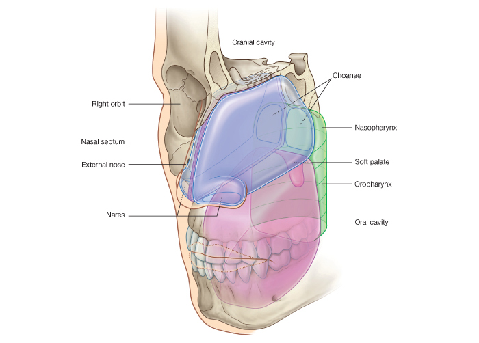
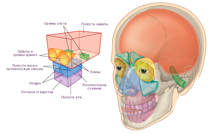
Схема. Главные отделы головы и шеи.
Модификация: Gray H., (1821–1865), Drake R., Vogl W., Mitchell A., Eds. Gray's Anatomy for Students. Churchill Livingstone, 2007, 1150 p., см.: Анатомия человека: Литература. Иллюстрации.
|
 |
|
|
Схема. Полости носа. А. Преддверие носа, крыша носа и боковые стенки носа. B. Раковины на боковых стенках носа. C. коронарное сечение. D. Воздушные каналы правой носовой полости = Nasal cavities. A. Floor, roof, and lateral walls. B. Conchae on lateral walls. C. Coronal section. D. Air channels in right nasal cavity.
Модификация: Gray H., (1821–1865), Drake R., Vogl W., Mitchell A., Eds. Gray's Anatomy for Students. Churchill Livingstone, 2007, 1150 p.
см.: Анатомия человека: Литература. Иллюстрации |
 |
|
|
Примечание:
В соответствии со структурой и функцией в слизистой оболочке полости носа выделяют обонятельную область и дыхательную область. |
 |
|
К обонятельной области относят часть слизистой оболочки носа содержащую обонятельные нейросенсорные клетки. Обонятельная область слизистой оболочки покрывает правую и левую верхние носовые раковины, часть средних носовых раковин, а также соответствующий их расположению верхний отдел перегородки носа. Остальная часть слизистой оболочки носа относится к дыхательной области.
Слизистая оболочка дыхательной области покрыта мерцательным эпителием. В ней содержатся слизистые и серозные железы.
В области нижней раковины слизистая оболочка и подслизистая основа содержат большое количество венозных сосудов. Эти сосуды образуют пещеристые венозные сплетения раковин. Наличие пещеристых венозных сплетений способствует согреванию вдыхаемого воздуха.
|
|
Примечание:
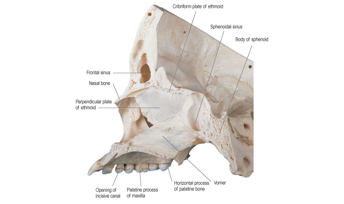
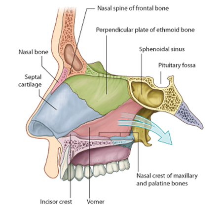
The medial wall of each nasal cavity is the mucosa-covered surface of the thin nasal septum, which is oriented vertically in the median sagittal plane and separates the right and left nasal cavities from each other. |
 |
|
The nasal septum consists of:
• the septal nasal cartilage anteriorly;
• posteriorly, mainly the vomer and the perpendicular plate of the ethmoid bone;
• small contributions by the nasal bones where they meet in the midline, and the nasal spine of the frontal bone;
• contributions by the crests of the maxillary and palatine bones, rostrum of the sphenoid bones, and the incisor crest of the maxilla.
|
|
Примечание:
The floor of each nasal cavity is smooth, concave, and much wider than the roof. It consists of:
• soft tissues of the external nose;
• the upper surface of the palatine process of the maxilla, and the horizontal plate of the palatine bone, which together form the hard palate.
The naris opens anteriorly into the floor, and the superior aperture of the incisive canal is deep to the mucosa immediately lateral to the nasal septum near the front of the hard palate. |
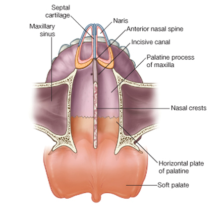 |
|
|
Примечание:
The roof of the nasal cavity is narrow and is highest in central regions where it is formed by the cribriform plate of the ethmoid bone.
Anterior to the cribriform plate the roof slopes inferiorly to the nares and is formed by:
• the nasal spine of the frontal bone and the nasal bones;
• the lateral processes of the septal cartilage and major alar cartilages of the external nose.
|
 |
|
Posteriorly, the roof of each cavity slopes inferiorly to the choana and is formed by:
• the anterior surface of the sphenoid bone;
• the ala of the vomer and adjacent sphenoidal process of the palatine bone; and
• the vaginal process of the medial plate of the pterygoid process.
Underlying the mucosa, the roof is perforated superiorly by openings in the cribriform plate, and anterior to these openings by a separate foramen for the anterior ethmoidal nerve and vessels.
The opening between the sphenoidal sinus and the spheno-ethmoidal recess is on the posterior slope of the roof.
|
|
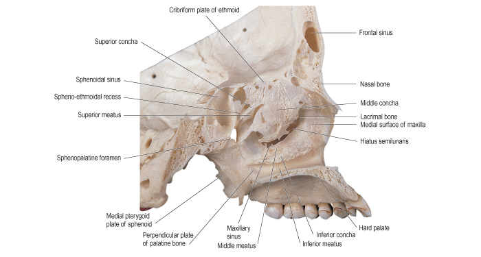
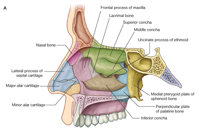
Схема. Боковая стенка полости носа. A. Кости = Lateral wall of the nasal cavity. A. Bones.
Модификация: Gray H., (1821–1865), Drake R., Vogl W., Mitchell A., Eds. Gray's Anatomy for Students. Churchill Livingstone, 2007, 1150 p.
см.: Анатомия человека: Литература. Иллюстрации |
 |
|
Примечание:
|
The lateral wall of each nasal cavity is complex and is formed by bone, cartilage, and soft tissues.
Bony support for the lateral wall (Fig. 8.226A) is provided by:
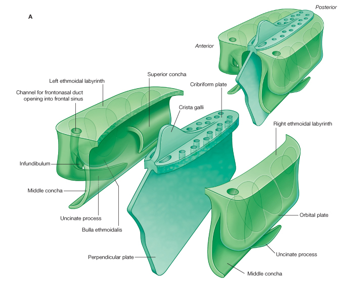
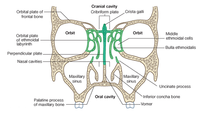
• the ethmoidal labyrinth and uncinate process;
• the perpendicular plate of the palatine bone;
• the medial plate of the pterygoid process of the sphenoid bone;
• the medial surfaces of the lacrimal bones and maxillae;
• the inferior concha.
In the external nose, the lateral wall of the cavity is supported by cartilage (lateral process of the septal cartilage and major and minor alar cartilages) and by soft tissues. The surface of the lateral wall is irregular in contour and is interrupted by the three nasal conchae.
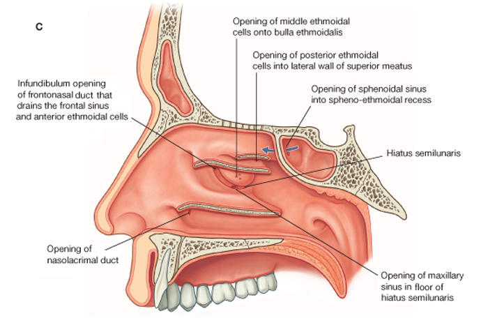
The inferior, middle, and superior conchae extend medially across the nasal cavity, separating it into four air channels, an inferior, middle, and superior meatus, and a spheno-ethmoidal recess. The conchae do not extend forward into the external nose. The anterior end of each concha curves inferiorly to form a lip that overlies the end of the related meatus.
Immediately inferior to the attachment of the middle concha and just anterior to the midpoint of the concha, the lateral wall of the middle meatus elevates to form the dome-shaped ethmoidal bulla. This is formed by the underlying middle ethmoidal cells, which expand the medial wall of the ethmoidal labyrinth.
Inferior to the ethmoidal bulla is a curved gutter (the hiatus semilunaris), which is formed by the mucosa covering the lateral wall as it spans a defect in the bony wall between the ethmoidal bulla above and the uncinate process below.
The anterior end of the hiatus semilunaris forms a channel (the ethmoidal infundibulum), which curves upwards and continues as the frontonasal duct through the anterior part of the ethmoidal labyrinth to open into the frontal sinus. |
|
|
Схема. Боковая стенка полости носа. C. Частично удалённые раковины, расположенные на боковых стенках полости носа = Lateral wall of the nasal cavity. C. Conchae broken away at attachment to lateral wall.
Модификация: Gray H., (1821–1865), Drake R., Vogl W., Mitchell A., Eds. Gray's Anatomy for Students. Churchill Livingstone, 2007, 1150 p.
см.: Анатомия человека: Литература. Иллюстрации |
 |
|
|
Примечание:
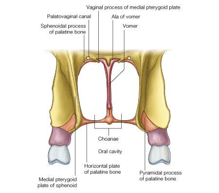
The choanae are the oval-shaped openings between the nasal cavities and the nasopharynx. |
 |
|
Unlike the nares, which have flexible borders of cartilage and soft tissues, the choanae are rigid openings completely surrounded by bone, and their margins are formed:
• inferiorly, by the posterior border of the horizontal plate of the palatine bone;
• laterally, by the posterior margin of the medial plate of the pterygoid process;
• medially, by the posterior border of the vomer. The roof of the choanae is formed:
• anteriorly by the ala of the vomer and the vaginal process of the medial plate of the pterygoid process;
• posteriorly by the body of the sphenoid bone.
|
|
Примечание:
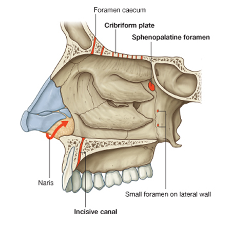
Gateways
There are a number of routes by which nerves and vessels enter and leave the soft tissues lining each nasal cavity, and these include the cribriform plate, sphenopalatine foramen, the incisive canal, small foramina in the lateral wall, and around the margin of the nares. |
 |
|
Cribriform plate
The fibers of the olfactory nerve [I] exit the nasal cavity and enter the cranial cavity through perforations in the cribriform plate. In addition, small foramina between the cribriform plate and surrounding bone allow the anterior ethmoidal nerve, a branch of the ophthalmic nerve [V1], and accompanying vessels to pass from the orbit into the cranial cavity and then down into the nasal cavity.
In addition, there is a connection in some individuals between nasal veins and the superior sagittal sinus of the cranial cavity through a prominent foramen (the foramen caecum) in the midline between the crista galli and frontal bone.
Sphenopalatine foramen
One of the most important routes by which nerves and vessels enter and leave the nasal cavity is the sphenopalatine foramen in the posterolateral wall of the superior nasal meatus. This foramen is just superior to the attachment of the posterior end of the middle nasal concha and is formed by the sphenopalatine notch in the palatine bone and the body of the sphenoid bone.
The sphenopalatine foramen is a route of communication between the nasal cavity and the pterygopalatine fossa. Major structures passing through the foramen are:
• the sphenopalatine branch of the maxillary artery;
• the nasopalatine branch of the maxillary nerve [V2];
• superior nasal branches of the maxillary nerve [V2].
Incisive canal
Another route by which structures enter and leave the nasal cavities is through the incisive canal in the floor of the nasal cavities. This canal is immediately lateral to the nasal septum and just posterosuperior to the root of the central incisor in the maxilla. It opens into the incisive fossa in the roof of the oral cavity and transmits:
• the nasopalatine nerve from the nasal cavity into the oral cavity;
• the terminal end of the greater palatine artery from the oral cavity into the nasal cavity.
Small foramina in the lateral wall
Other routes by which vessels and nerves get into and out of the nasal cavity include the nares and small foramina in the lateral wall:
• internal nasal branches of the infra-orbital nerve of the maxillary nerve [V2] and alar branches of the nasal artery from the facial artery
• loop around the margin of the naris to gain entry to the lateral wall of the nasal cavity from the face;
• inferior nasal branches from the greater palatine branch of the maxillary nerve [V2] enter the lateral wall of the nasal cavity from the palatine canal by passing through small foramina on the lateral wall.
|
|
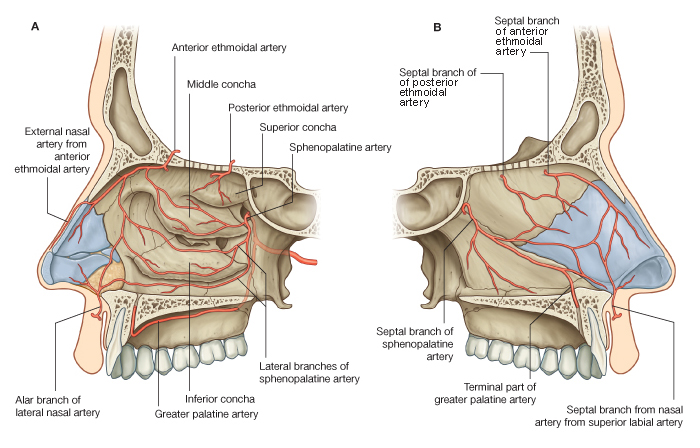
Схема. Артерии полостей носа. A. Боковая стенка правой половины носовой полости. B. Перегородка носовой полости (медиальная стенка правой половины носовой полости) = Arterial supply of the nasal cavities. A. Lateral wall of the right nasal cavity. B. Septum (medial wall of right nasal cavity).
Модификация: Gray H., (1821–1865), Drake R., Vogl W., Mitchell A., Eds. Gray's Anatomy for Students. Churchill Livingstone, 2007, 1150 p.
см.: Анатомия человека: Литература. Иллюстрации |
 |
|
|
Примечание:
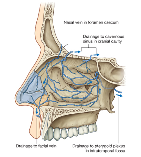
Veins draining the nasal cavities generally follow the arteries:
• veins that pass with branches that ultimately originate from the maxillary artery drain into the pterygoid plexus of veins in the infratemporal fossa;
• veins from anterior regions of the nasal cavities join the facial vein. |
 |
|
In some individuals, an additional nasal vein passes superiorly through a midline aperture (the foramen caecum), in the frontal bone anterior to the crista galli, and joins with the anterior end of the superior sagittal sinus. Because this nasal vein connects an intracranial venous sinus with extracranial veins, it is classified as an emissary vein. Emissary veins in general are routes by which infections can track from peripheral regions into the cranial cavity.
Veins that accompany the anterior and posterior ethmoidal arteries are tributaries of the superior ophthalmic vein, which is one of the largest emissary veins and drains into the cavernous sinus on either side of the hypophysial fossa.
|
|
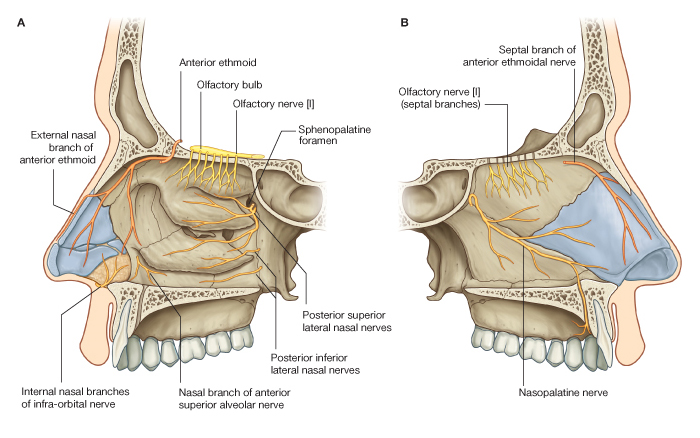
Схема. Иннервация полостей носа. A. Боковая стенка правой полости носа. B. Медиальная стенка правой полости носа = Innervation of the nasal cavities. A. Lateral wall of right nasal cavity. B. Medial wall of right nasal cavity.
Модификация: Gray H., (1821–1865), Drake R., Vogl W., Mitchell A., Eds. Gray's Anatomy for Students. Churchill Livingstone, 2007, 1150 p.
см.: Анатомия человека: Литература. Иллюстрации |
 |
|
Примечание:
|
Nerves that innervate the nasal cavities are:
• the olfactory nerve [I] for olfaction;
• branches of the ophthalmic [V1] and maxillary [V2] nerves for general sensation.
Secretomotor innervation of mucous glands in the nasal cavities and paranasal sinuses is by parasympathetic fibers from the facial nerve [VII], which mainly join branches of the maxillary nerve [V2] in the pterygopalatine fossa.
Olfactory nerve [I]
The olfactory nerve [I] is composed of axons from receptors in the olfactory epithelium at the top of each nasal cavity. Bundles of these axons pass superiorly through perforations in the cribriform plate to synapse with neurons in the olfactory bulb of the brain.
Branches from the ophthalmic nerve [V1]
Branches from the ophthalmic nerve [V1] that innervate the nasal cavity are the anterior and posterior ethmoidal nerves, which originate from the nasociliary nerve in the orbit.
Anterior and posterior ethmoidal nerves
The anterior ethmoidal nerve (Fig. 8.232) travels with the anterior ethmoidal artery and leaves the orbit through a canal between the ethmoidal labyrinth and the frontal bone. It passes through and supplies the adjacent ethmoidal cells and frontal sinus, and then enters the cranial cavity immediately lateral and superior to the cribriform plate.
The anterior ethmoidal nerve travels forward in a groove on the cribriform plate and then enters the nasal cavity by descending through a slit-like foramen immediately lateral to the crista galli. It has branches to the medial and lateral wall of the nasal cavity and then continues forward on the undersurface of the nasal bone. It passes onto the external surface of the nose by traveling between the nasal bone and lateral nasal cartilage, and then terminates as the external nasal nerve, which supplies skin around the naris, in the nasal vestibule, and on the tip of the nose.
Like the anterior ethmoidal nerve, the posterior ethmoidal nerve leaves the orbit through a similar canal in the medial wall of the orbit. It terminates by supplying the mucosa of the ethmoidal cells and sphenoidal sinus and normally does not extend into the nasal cavity itself.
Branches from the maxillary nerve [V2]
A number of nasal branches from the maxillary nerve [V2] innervate the nasal cavity. Many of these nasal branches (Fig. 8.232) originate in the pterygopalatine fossa, which is just lateral to the lateral wall of the nasal cavity, and leave the fossa to enter the nasal cavity by passing medially through the sphenopalatine foramen:
• a number of these nerves (posterior superior lateral nasal nerves) pass forward on and supply the lateral wall of the nasal cavity;
• others (posterior superior medial nasal nerves) cross the roof to the nasal septum and supply both these regions;
• the largest of these nerves is the nasopalatine nerve, which passes forward and down the medial wall of the nasal cavity to pass through the incisive canal onto the roof of the oral cavity, and terminates by supplying the oral mucosa posterior to the incisor teeth;
• other nasal nerves (posterior inferior nasal nerves) originate from the greater palatine nerve, descending from the pterygopalatine fossa in the palatine canal just lateral to the nasal cavity, and pass through small bony foramina to innervate the lateral wall of the nasal cavity;
• a small nasal nerve also originates from the anterior superior alveolar branch of the infra-orbital nerve and passes medially through the maxilla to supply the lateral wall near the anterior end of the inferior concha.
Parasympathetic innervation
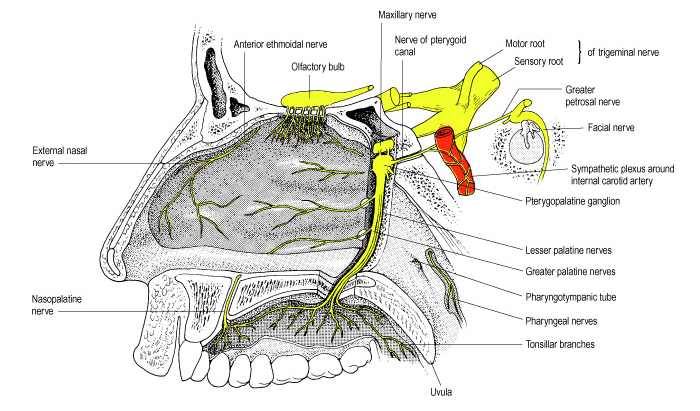
Secretomotor innervation of glands in the mucosa of the nasal cavity and paranasal sinuses is by preganglionic parasympathetic fibers carried in the greater petrosal branch of the facial nerve [VII]. These fibers enter the pterygopalatine fossa and synapse in the pterygopalatine ganglion (see p. 891). Postganglionic parasympathetic fibers then join branches of the maxillary nerve [V2] to leave the fossa and ultimately reach target glands.
Sympathetic innervation
Sympathetic innervation, mainly involved with regulating blood flow in the nasal mucosa, is from the spinal cord level T1. Preganglionic sympathetic fibers enter the sympathetic trunk and ascend to synapse in the superior sympathetic ganglion. Postganglionic sympathetic fibers pass onto the internal carotid artery, enter the cranial cavity, and then leave the internal carotid artery to form the deep petrosal nerve, which joins the greater petrosal nerve of the facial nerve [VII] and enters the pterygopalatine fossa (see Fig. 8.147 and p. 896).
Like the parasympathetic fibers, the sympathetic fibers follow branches of the maxillary nerve [V2] into the nasal cavity. |
|
|
Примечание:
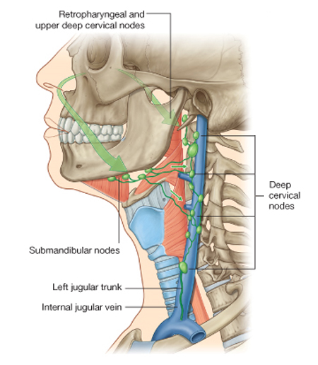
Lymph from anterior regions of the nasal cavities drains forward onto the face by passing around the margins of the nares (Fig. 8.233). These lymphatics ultimately connect with the submandibular nodes.
Lymph from posterior regions of the nasal cavity and the paranasal sinuses drains into upper deep cervical nodes. Some of this lymph passes first through the retropharyngeal nodes.
|
 |
|
|
Схема. Артерии боковой стенки полости носа = Arteries of the lateral wall of the nose.
Модификация: Gray H., (1821–1865), Drake R., Vogl W., Mitchell A., Eds. Gray's Anatomy for Students. Churchill Livingstone, 2007, 1150 p., см.: Анатомия человека: Литература. Иллюстрации.
|
 |
|
|
Схема. Сенсорная иннервация боковой стенки полости носа, твёрдого и мягкого нёба и носоглотки = The sensory innervation of the lateral wall of the nasal cavity, hard and soft palates, and nasopharynx.
Модификация: Gray H., (1821–1865), Drake R., Vogl W., Mitchell A., Eds. Gray's Anatomy for Students. Churchill Livingstone, 2007, 1150 p., см.: Анатомия человека: Литература. Иллюстрации.
|
 |
|
Примечание:
|
Secretomotor fibres to mucous glands are distributed in branches from the pterygopalatine ganglion. |
|
|
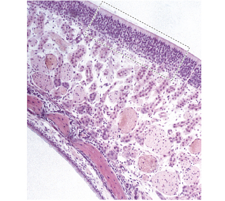
Схема. Микрофотография обонятельной области слизистой оболочки носа, покрывающей верхние раковины (низкое разрешение) = Low power micrograph of the olfactory mucosa covering the superior concha.
Модификация: Gray H., (1821–1865), Standring S., Ed. Gray's Anatomy: The Anatomical Basis of Clinical Practice. 39th ed., Churchill Livingstone, 2008, 1600 p.
см.: Анатомия человека: Литература. Иллюстрации.
Примечание:
The olfactory epithelium is shown overlying a lamina propria containing cavernous vascular tissue and olfactory glands (of Bowman). Bundles of olfactory axons (fila olfactoria) pass through the mucosa towards the cribriform plate. A thinner respiratory epithelium covers the lower surface, shown here beneath the bone of the concha. |
 |
|
Olfactory epithelium
The peripheral receptors for olfactory sensation are located bilaterally in areas of sensory epithelium lining the posterodorsal parts of the nasal cavities. The sensory epithelium occupies an area of c.5 cm2 covering the posterior upper parts of the lateral nasal wall, including the back of the superior concha, the sphenoethmoidal recess, the upper part of the perpendicular plate of the ethmoid and the roof of the nose arching between the septum and lateral wall, including the underside of the cribriform plate. This area is pigmented yellowish brown in contrast to the pinker colour of the respiratory mucosa.
Microstructure of the olfactory mucosa
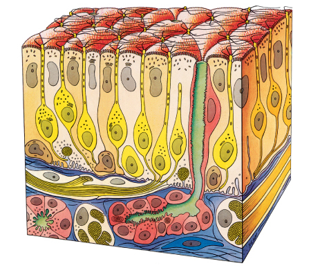
The olfactory mucosa consists of a pseudostratified olfactory epithelium, derived from the embryonic olfactory placodes (p. 245). It contains sensory receptor neurones, and its underlying lamina propria contains their axons, which lie within numerous olfactory nerve fascicles, and subepithelial olfactory glands (of Bowman). The glands secrete a predominantly serous fluid through ducts which open onto the epithelial surface. Their secretions form a thin fluid layer in which sensory cilia and the microvilli of sustentacular cells are embedded.
Olfactory epithelium
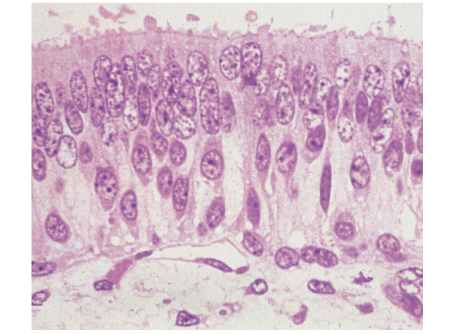
The olfactory epithelium (Figs 32.5, 32.6) is considerably thicker (up to 100 ?m) than the respiratory epithelium. It contains olfactory receptor neurones, sustentacular cells and two classes of basal cell, horizontal basal cells (closest to the basal lamina) and globose basal cells (Fig. 32.7). The nuclei of these various cells occupy specific zones within the epithelial thickness. Most superficially is a layer of sustentacular cell nuclei; beneath this, and occupying much of the epithelial thickness, are several tiers of receptor cell bodies and nuclei. Basal cells lie between this zone and the basal lamina underlying the epithelium.
|
|
Примечание:
Showing the nuclei of basal cells, olfactory neurones and sustentacular cells. The olfactory neuronal endings (knobs or vesicles) can be seen projecting from the free surface. |
 |
|
Olfactory epithelium
The peripheral receptors for olfactory sensation are located bilaterally in areas of sensory epithelium lining the posterodorsal parts of the nasal cavities. The sensory epithelium occupies an area of c.5 cm2 covering the posterior upper parts of the lateral nasal wall, including the back of the superior concha, the sphenoethmoidal recess, the upper part of the perpendicular plate of the ethmoid and the roof of the nose arching between the septum and lateral wall, including the underside of the cribriform plate. This area is pigmented yellowish brown in contrast to the pinker colour of the respiratory mucosa.
Microstructure of the olfactory mucosa
The olfactory mucosa consists of a pseudostratified olfactory epithelium, derived from the embryonic olfactory placodes (p. 245). It contains sensory receptor neurones, and its underlying lamina propria contains their axons, which lie within numerous olfactory nerve fascicles, and subepithelial olfactory glands (of Bowman). The glands secrete a predominantly serous fluid through ducts which open onto the epithelial surface. Their secretions form a thin fluid layer in which sensory cilia and the microvilli of sustentacular cells are embedded.
Olfactory epithelium
The olfactory epithelium (Figs 32.5, 32.6) is considerably thicker (up to 100 ?m) than the respiratory epithelium. It contains olfactory receptor neurones, sustentacular cells and two classes of basal cell, horizontal basal cells (closest to the basal lamina) and globose basal cells (Fig. 32.7). The nuclei of these various cells occupy specific zones within the epithelial thickness. Most superficially is a layer of sustentacular cell nuclei; beneath this, and occupying much of the epithelial thickness, are several tiers of receptor cell bodies and nuclei. Basal cells lie between this zone and the basal lamina underlying the epithelium.
|
|
Примечание:
Receptor cells (yellow) are situated among columnar sustentacular cells. The axons of the receptor cells emerge from the epithelium in bundles enclosed by ensheathing glial cells. Rounded globose basal cells (brown) and flattened horizontal basal cells (not shown) lie on the basal lamina and the subepithelial glands (of Bowman) open on to the surface via their intraepithelial ducts (green). At the surface are cilia of the receptor cells and microvilli of the supporting cells. |
 |
|
Olfactory receptor neurones are bipolar cells. They have a cell body and nucleus located in the middle zone of the epithelium, a single unbranched apical dendrite c.2 ?m across which extends to the epithelial surface, and a basally directed unmyelinated axon c.0.2 ?m in diameter, which passes out of the epithelium. Each dendrite projects into the overlying secretory fluid as an expanded cylindrical olfactory ending (rod, knob or vesicle). Groups of up to 20 cilia radiate from the circumference of each ending and extend for long distances parallel to the epithelial surface. Internally, the short proximal part of each cilium has the '9 + 2' pattern of microtubules typical of motile cilia (p. 19), while the longer distal trailing end contains only the central pair of microtubules. The cilia lack dynein arms on the peripheral microtubule doublets and are thought to be non-motile, serving to project a large area of sensory surface for the efficient detection of odorants. Individual receptor neurones express receptors for a single (or very few) odorant molecules. Although neurones with the same receptor specificity are randomly distributed within anatomical zones of the epithelium, all project to the same target dendritic field (glomeruli) of the olfactory bulb, and there is a considerable degree of convergence. Specific odours activate a unique spectrum of receptors which in turn activate restricted groups of glomeruli and their second order neurones.
The axons form small intraepithelial fascicles among the processes of sustentacular and basal cells. The fascicles penetrate the basal lamina, and are immediately ensheathed by olfactory ensheathing cells. Groups of up to 50 fascicles join to form larger olfactory nerve rootlets which pass through the cribriform plate to enter the olfactory bulb, there synapsing in glomeruli with secondary sensory neurones, principally mitral cells and, to a lesser extent, smaller tufted cells.
Sustentacular cells
Sustentacular, or supporting, cells (Fig. 32.7) are columnar cells that separate and partially ensheathe the olfactory receptors. Their large nuclei form a layer superficial to the receptor nuclei. They send numerous long, irregular microvilli into the secretory fluid layer covering the surface of the epithelium, where they lie among the long trailing ends of olfactory receptor cilia. At their bases, facing the basal lamina, they have expanded end-feet containing numerous lamellated dense bodies resembling neuronal lipofuscin granules. These are remains of secondary lysosomes formed as a result of phagocytic activity, and are largely responsible for the pigmentation of the olfactory area. The granules gradually accumulate with age, and because these cells are long-lived, pigmentation also increases in intensity with age. Cells are linked by desmosomes near the epithelial surface, which gives mechanical coherence to the epithelium, and by tight junctions between the sustentacular cells and olfactory receptors at the level of the epithelial surface, which provides a protective barrier.
Basal cells
There are two types of basal cell; horizontal basal cells and globose basal cells. Horizontal basal cells are flattened against the basal lamina, and have condensed nuclei and darkly staining cytoplasm containing numerous intermediate filaments of the cytokeratin family, inserted into desmosomes contacting surrounding sustentacular cells. In contrast, globose cells are rounded or elliptical in shape, with a pale, euchromatic nucleus, and a pale cytoplasm. They form a distinct zone spaced slightly from the basal surface of the epithelium. Mitoses are found within this zone because globose basal cells are the immediate source of new olfactory receptor neurones.
Olfactory ensheathing (glial) cells
Olfactory ensheathing cells share properties with astrocytes and non-myelinating Schwann cells, but they possess distinctive features that indicate they are a separate class of glia. Developmentally they are derived from the olfactory placode rather than neural crest. They ensheath olfactory axons in a unique manner throughout their entire course, and accompany them into the central nervous tissue of the olfactory bulb, where they contribute to the glia limitans.
Olfactory glands
The olfactory (Bowman's) glands (Fig. 32.7) are branched tubuloalveolar structures that lie beneath the olfactory epithelium and secrete onto the epithelial surface through narrow, vertical ducts. Their secretions, which include defensive substances, lysozyme, lactoferrin, IgA and sulphated proteoglycans, bathe the dendritic endings and cilia of the olfactory receptors. The fluid acts as a solvent for odorant molecules, allowing their diffusion to the sensory receptors. The glands also secrete odorant-binding proteins into the fluid, which increase the efficiency of odour detection.
Turnover of olfactory receptor neurons
Receptor neurones are lost and replaced throughout life. Individual receptor cells have a variable lifespan, averaging 1-3 months. Stem cells situated near the base of the epithelium undergo periodic mitotic division throughout life, giving rise to new olfactory receptor cells which then grow a dendrite to the olfactory surface and an axon to the olfactory bulb. The cell bodies of these new receptor neurones gradually move apically until they reach the region just below the supporting cell nuclei. When they degenerate, dead cells are either shed from the epithelium or are phagocytosed by sustentacular cells. The rate of receptor cell loss and replacement increases after exposure to damaging stimuli, but their capacity to turnover declines slowly with age, and this contributes to diminishing olfactory sensory function in old age.
|
|
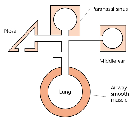
Схема. Непосредственные взаимоотношения между полостями рта, носа, синусов, среднего уха и лёгких = Intimate connections between the nose, the sinuses, the middle ear and the lungs.
Модификация: Scadding G.K., Lund V.J., Eds. Investigative Rhinology, Taylor & Francis, 2004, 208 p., см.: Физиология человека: Литература. Иллюстрации.
Примечание:
Rhinitis has significant comorbid associations related to its intimate
connections between the nose, the sinuses, the middle ear and
the lungs. These include asthma, sinusitis, pharyngitis,
otitis media with effusion and sleep problems. Most is known about
the rhinitis–asthma link.
|
 |
|
The mechanisms of interaction between the nose and chest are
not fully elucidated – certainly the ciliated pseudocolumnar respiratory
epithelium is continuous from just inside the nose to the
smaller bronchi and is likely to react in similar fashion all along its
length. Other factors probably include nasal obstruction leading to mouth breathing and loss of the normal filtration, warming and
humidification of inspired air. The passage of postnasal secretions
containing inflammatory or infectious mediators is unlikely to
cause significant lower respiratory problems since it normally passes
into the posterior pharynx and is swallowed rather than being
inhaled. Naso-sino-bronchial reflexes exist; systemic connections between the nose and lungs involving the production and release
of eosinophil precursors bearing high affinity IL-5 (interleukin-5)
receptors have been demonstrated. Nitric oxide, predominantly
produced in the sinuses and upper respiratory tract, may also be
relevant.
|
Литература. Иллюстрации. References. Illustrations
Щелкни здесь и получи доступ в библиотеку сайта! Click here and receive access to the reference library!
- Бабияк В.И., Накатис Я.А. Клиническая оториноларингология, СПб., Гиппократ, 2005, 795 с.
Учебное пособие.
Доступ к данному источнику = Access to the reference.
URL: http://www.tryphonov.ru/tryphonov/serv_r.htm#0 quotation
- Воячек В.И. Военная оториноларингология, М., Медгиз, 1946, 150 с.
Учебное пособие.
Доступ к данному источнику = Access to the reference.
URL: http://www.tryphonov.ru/tryphonov/serv_r.htm#0 quotation
- Компанеец С.М., Скрыпт А.А., Ред. Болезни уха, носа и горла. Киев, Государственное медицинское издательство УССР, 1941 с. Трехтомное руководство для врачей, подготовленное большим коллективом специалистов.
Цитата из данного источника. Формат .CHM.
URL: http://www.tryphonov.ru/tryphonov/serv_r.htm#0 quotation
- Лихачев А.Г., ред. Руководство по оториноларингологии. Анатомия, физиология, эмбриология и методы исследования, М., Гос.изд-во мед. лит., 1960, 640 с.
Учебное пособие.
Доступ к данному источнику = Access to the reference.
URL: http://www.tryphonov.ru/tryphonov/serv_r.htm#0 quotation
- Пальчун В.Т., Крюков А.И. Оториноларингология: Руководство для врачей. М.: «Медицина», 2001, 616 с. Руководство.
Доступ к данному источнику = Access to the reference.
URL: http://www.tryphonov.ru/tryphonov/serv_r.htm#0 quotation
- Avery J.K., Ed. Oral Development and Histology = Гистология и развитие полости рта. 3rd ed., Thieme, 2001, 460 p.
Иллюстрированное учебное пособие. .
Доступ к данному источнику = Access to the reference.
URL: http://www.tryphonov.ru/tryphonov/serv_r.htm#0 quotation
- Berkovitz B.K.B., Moxham B.J., Linden R.W.A., Sloan A.J., Eds. Oral Biology: Oral Anatomy, Histology, Physiology and Biochemistry = Анатомия, гистология, физиология и биохимия органов полости рта, Elsevier, 2011, 311 p.
Иллюстрированное учебное пособие. .
Доступ к данному источнику = Access to the reference.
URL: http://www.tryphonov.ru/tryphonov/serv_r.htm#0 quotation
- Cawson R.A., Odell J.W. Essentials of Oral Pathology and Oral Medicine = Основы патологии и медицины полости рта. Churchill Livingstone, 2002, 385 p. Иллюстрированный справочник.
Доступ к данному источнику = Access to the reference.
URL: http://www.tryphonov.ru/tryphonov/serv_r.htm#0 quotation
- Dayan N., Wertz W., Eds. Innate Immune System of Skin and Oral Mucosa: Properties and Impact in Pharmaceutics, Cosmetics, and Personal Care Products = Врождённая иммунная система кожи и слизистой полости рта. Свойства и неблагоприятные влияния фармакологических препаратов, косметики и индивидуальных средств ухода. Wiley, 2011, 351 p. Обзоры.
Доступ к данному источнику = Access to the reference.
URL: http://www.tryphonov.ru/tryphonov/serv_r.htm#0 quotation
Учебное пособие. .
Цитата из данного источника.
URL: http://www.tryphonov.ru/tryphonov/serv_r.htm#0 quotation
- Field A., Longman L., Tyldesley W.R., Eds. Tyldesley's Oral Medicine = Стоматология. 5th ed., Oxford University Press, 2003, 240 p.Учебное пособие. .
Доступ к данному источнику = Access to the reference.
URL: http://www.tryphonov.ru/tryphonov/serv_r.htm#0 quotation
- National Academy of Sciences. Oral Health Literacy = Грамотность в области гигиены полости рта. National Academy of Sciences, 2013, 143 p.
Популярная брошюра.
Доступ к данному источнику = Access to the reference.
URL: http://www.tryphonov.ru/tryphonov/serv_r.htm#0 quotation
- Scadding G.K., Lund V.J. Investigative Rhinology = Исследования в ринологии. Informa HealthCare, 2004, 208 p. Иллюстрированное учебное пособие. .
Доступ к данному источнику = Access to the reference.
URL: http://www.tryphonov.ru/tryphonov/serv_r.htm#0 quotation
- Sterer N., Rosenberg M. Breath Odors: Origin, Diagnosis, and Management = Дурной запах изо рта. Происхождение, диагностика, устранение. Springer-Verlag Berlin Heidelberg 2011 , 112 p.
Иллюстрированное учебное пособие. .
Доступ к данному источнику = Access to the reference.
URL: http://www.tryphonov.ru/tryphonov/serv_r.htm#0 quotation
- Ten Cate’s Oral Histology: Development, Structure, and Function = Гистология полости рта. 6th ed., Mosby, 2003.
Программа. Иллюстрированное учебное пособие. .
Доступ к данному источнику = Access to the reference.
URL: http://www.tryphonov.ru/tryphonov/serv_r.htm#0 quotation
|
«Я У Ч Е Н Ы Й И Л И . . . Н Е Д О У Ч К А ?»
Т Е С Т В А Ш Е Г О И Н Т Е Л Л Е К Т А
Предпосылка:
Эффективность развития любой отрасли знаний определяется степенью соответствия методологии познания - познаваемой сущности.
Реальность:
Живые структуры от биохимического и субклеточного уровня, до целого организма являются вероятностными структурами. Функции вероятностных структур являются вероятностными функциями.
Необходимое условие:
Эффективное исследование вероятностных структур и функций должно основываться на вероятностной методологии (Трифонов Е.В., 1978,..., ..., 2015, …).
Критерий: Степень развития морфологии, физиологии, психологии человека и медицины, объём индивидуальных и социальных знаний в этих областях определяется степенью использования вероятностной методологии.
Актуальные знания: В соответствии с предпосылкой, реальностью, необходимым условием и критерием...
...
о ц е н и т е с а м о с т о я т е л ь н о:
— с т е п е н ь р а з в и т и я с о в р е м е н н о й н а у к и,
— о б ъ е м В а ш и х з н а н и й и
— В а ш и н т е л л е к т !
|
♥ Ошибка? Щелкни здесь и исправь ее! Поиск на сайте E-mail автора (author): tryphonov@yandex.ru
|

